ultrasound Scan
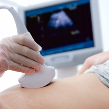
Ultrasound Scan is a safe non invasive, cost effective & almost accurate investigation. It also plays an important role in the care of every pregnant woman. State-of-the-art technology at Dr Joy scan Fetal Assessment Center allows for early diagnosis, further analysis and treatment plans.
Ultrasound Scan Services At Joy Scan & Diagnostic Center
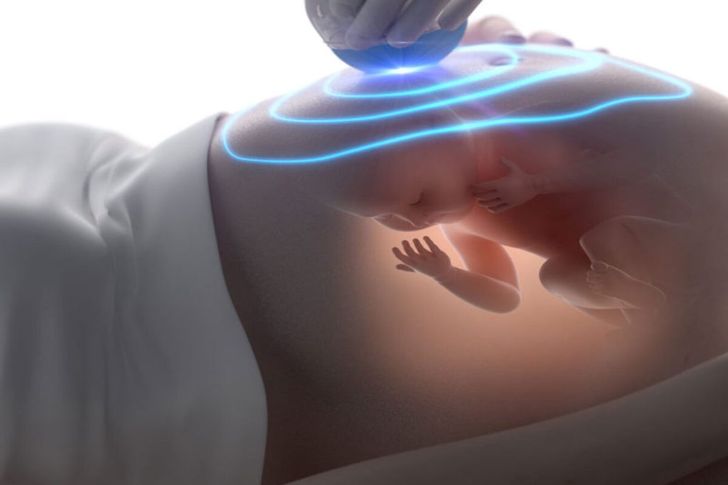
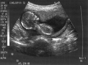
FOETAL SCANS INCLUDING FOETAL 2D ECHO
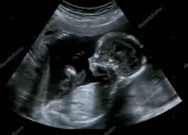
A Fetal ultrasound (sonogram/sonography) is an imaging technique that uses sound waves to produce images of a fetus in the uterus

MALE FEMALE & TRANSGENDER ABDOMINAL SCANS
An Abdominal Scan is a painless test that uses a specialized X-ray machine to take pictures of a patient's organs, blood vessels and lymph nodes.
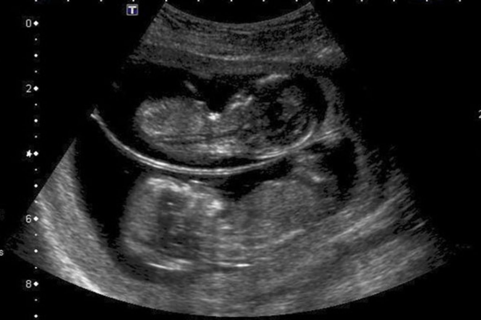
MALE & FEMALE INFERTILITY SCANS
Infertility is the inability of a person to reproduce by natural means. It is usually not the natural state of a healthy adult, except notably among certain social species
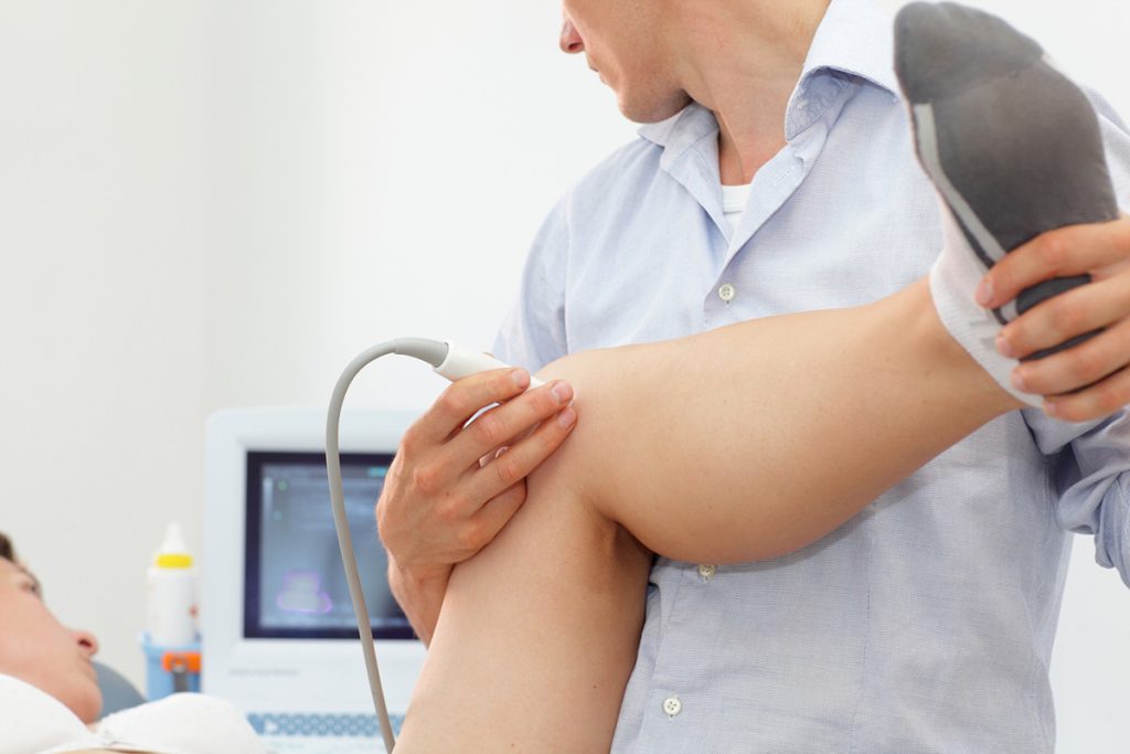
MUSCULO SKELETAL SCANS
Ultrasound - Musculoskeletal Ultrasound imaging uses sound waves to produce pictures of muscles, tendons, ligaments, nerves and joints throughout the body
– Dating & Viability Scan
– Nuchal Translucency Scan At 11-14 Weeks
– 1st Trimester Soft Marker Genetic Scan
– Scan When Unable To Feel Foetal Movements For Foetal Well Being
– Triple Screening Scan
– Anamoly Scan – Best At 18- 24 Weeks
– 3d/4d Scan- Bonding Scan
– Foetal 2decho
– Maternal Uterine Artery Doppler Study – This Study Is During 21-23 Weeks
– Cervical Length Assessement Scan – 14 Weeks & 22 Weeks
– Foetal Doppler Studies – 24 Weeks Onwards
– Foetal Growth Assesement For Diagnosing LUGR
Screening tests for Down’s syndrome
– 11-14 weeks
– Nuchal translucency (NT)
– Combined test (NT, hCG, and PAPP-A)
– 14-20 weeks
– Triple test (hCG, AFP, and uE3)
– Quadruple test (hCG, AFP, uE3, and inhibin A)
– 11-14 weeks and 14-20 weeks
– Integrated test (NT, PAPP-A, inhibin A, hCG, AFP, and uE3)
– Serum integrated tests (PAPP-A, inhibin A, hCG, AFP, and uE3
– Detection & Exclusion Of Gall Stones
– Assess Liver For Size Shape And Condition
– Pancreas in normal, diabetes, alcohol
– Spleen & Central Blood Vessels
– Renal & Lower Abdominal Pain
– Abdominal Aorta Its Caliber & Flow Pattern
– Evaluate appendix
– Bowel loops , lymph nodes etc
Young girls – Unattainable puberty
– Unusual bleeding
– Pelvic pain
– Missed period scan
– Fertility & infertility
– EC Topic pregnancy
– Lesbian couple
– Tubal Patency
– Evaluate Uterus & Ovaries Prior To Conception
– Estimated Time Of Ovulation
– Measure Follicles & Endometrial Thickness before IVF
– Uterine Biophysical Prof

– Unusual Pain
– Unusual Bleeding
– Endometriosis
– Intrauterine Contraceptive Device (IUCD) & Mirena
– Heaviness Lower Abdomen
– Well Women Scan
– Family Planning
– Incontinence
– Prolapse
– Post Hysterectomy Pain
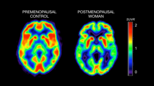
– Menopause
– Post Menopause
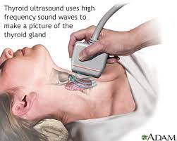
– Thyroid scan
– Breast scan
– Scrotal scan
– Trans rectal scans for prostate
– Any soft tissue swelling

– Shoulder scan for rotatory cuff, knee scan
– Ankle scan to evaluate Achilles tendon
– Sub-cutaneous scan
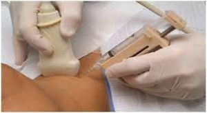
– Guided F N A C
– Percutaneous
– Transhepatic biliary drainage, Drainage of abscess
– Post surgical, Tubo-ovarian,hepatic, Appendicular, Renal, Retroperitoneal abscess
– Bakers cyst, Joint effusion drainage
– Breast abscess / Nodule aspiration
– Thoracentesis
– Amnioinfusion
– Ascitic fluid aspiration
– Prostate biopsy
– Minimally invasive focal tissue ablation
– Drainage of ganglion cysts
– Pain management
– Catheter guided thrombolytic therapy in veins & arteries (to save limb from reduced supply due to clot in them)
– Varicose vein ablation by laser/ Radiofrequency ablation
– Image guided insertion of central lines
– IVC filter insertion
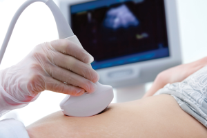
*Early pregnancy:1st trimester 5-14 weeks – TVS / TAS
*Confirmation of pregnancy
*Viability scan
*Evaluate the reason for spotting
*11 weeks Interval growth rate scan
*13 weeks Nuchal translucency scan
*1st-trimester soft marker scan
*Cervical length assessment scan
*14 weeks of cardiac evaluation
*Pre-eclampsia screening scan
*Evaluation of the nasal bone & presence of tricuspid flow for evidence of regurgitation
*Early foetal anatomical survey scan- EPAS
*Growth scan & double screening
*Placenta evaluation
*Site position thickness vascularisation volume to evaluate adverse *Pregnancy outcome & as a predictor in IUGR development
*Assessment of placenta in BOH -:Placental volume and vascularization index (VI), flow index (FI), and vascularization flow index (VFI) calculation to relate pregnancy outcomes
*Umbilical cord insertion level
*2nd trimester: 14 – 24 weeks:
*16-18 weeks Foetal anatomical survey scan (23 points evaluated)
*24 weeks Foetal echo
*2nd-trimester soft marker scan Second-trimester evaluation of cervical length for prediction of spontaneous preterm birth
**Foetal functional evaluation & foetal environmental studies
*15 – 21 weeks scan for triple marker screening Screening for Cardiac *Anomalies During the Early Second Trimester (as part of the *Triple/combined Test Ultrasound
*Prediction of preeclampsia by uterine artery colour Doppler velocimetry
*3rd trimester 24- 40 weeks
*Late anomaly scan
*28 – 30 weeks Interval growth rate scan
*32 – 36 weeks Feto placental Doppler studies. Foetal biophysical profile
*EPAS: Early pregnancy anomaly scan
*FASTER: First- and Second-Trimester Evaluation of Risk of Down’s Syndrome RADIUS
*Routine Antenatal Diagnostic Imaging with Ultrasound Study: TIFFA: *Targeted Imaging for foetal anomalies
*Amnioinfusion for the prevention of meconium aspiration syndrome
