FOETAL SCANS

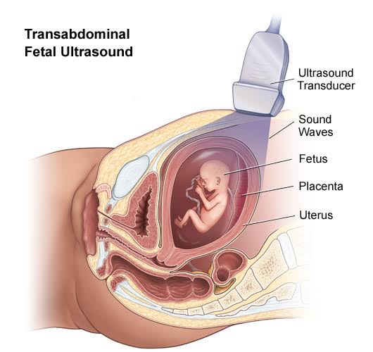
Obstetric ultrasonography, or prenatal ultrasound, is the use of
medical ultrasonography in pregnancy, in which sound waves are used to
create real-time visual images of the developing embryo or fetus in the
uterus (womb). The procedure is a standard part of prenatal care in many
countries, as it can provide a variety of information about the health
of the mother, the timing and progress of the pregnancy, and the health
and development of the embryo or fetus.
Scanning Procedure:
–Dating & Viability Scan
– Nuchal Translucency Scan At 11-14 Weeks
– 1st Trimester Soft Marker Genetic Scan
– Scan When Unable To Feel Foetal Movements For Foetal Well Being
– Triple Screening Scan
– Anamoly Scan – Best At 18- 24 Weeks
– 3d/4d Scan- Bonding Scan
– Foetal 2decho
– Maternal Uterine Artery Doppler Study – This Study Is During 21-23 Weeks
– Cervical Length Assessement Scan – 14 Weeks & 22 Weeks
– Foetal Doppler Studies – 24 Weeks Onwards
– Foetal Growth Assesement For Diagnosing LUGR
Screening tests for Down’s syndrome
– 11-14 weeks
– Nuchal translucency (NT)
– Combined test (NT, hCG, and PAPP-A)
– 14-20 weeks
– Triple test (hCG, AFP, and uE3)
– Quadruple test (hCG, AFP, uE3, and inhibin A)
– 11-14 weeks and 14-20 weeks
– Integrated test (NT, PAPP-A, inhibin A, hCG, AFP, and uE3)
– Serum integrated tests (PAPP-A, inhibin A, hCG, AFP, and uE3
